KIDNEY AND URINARY TRACT STONES
Kidney Stones
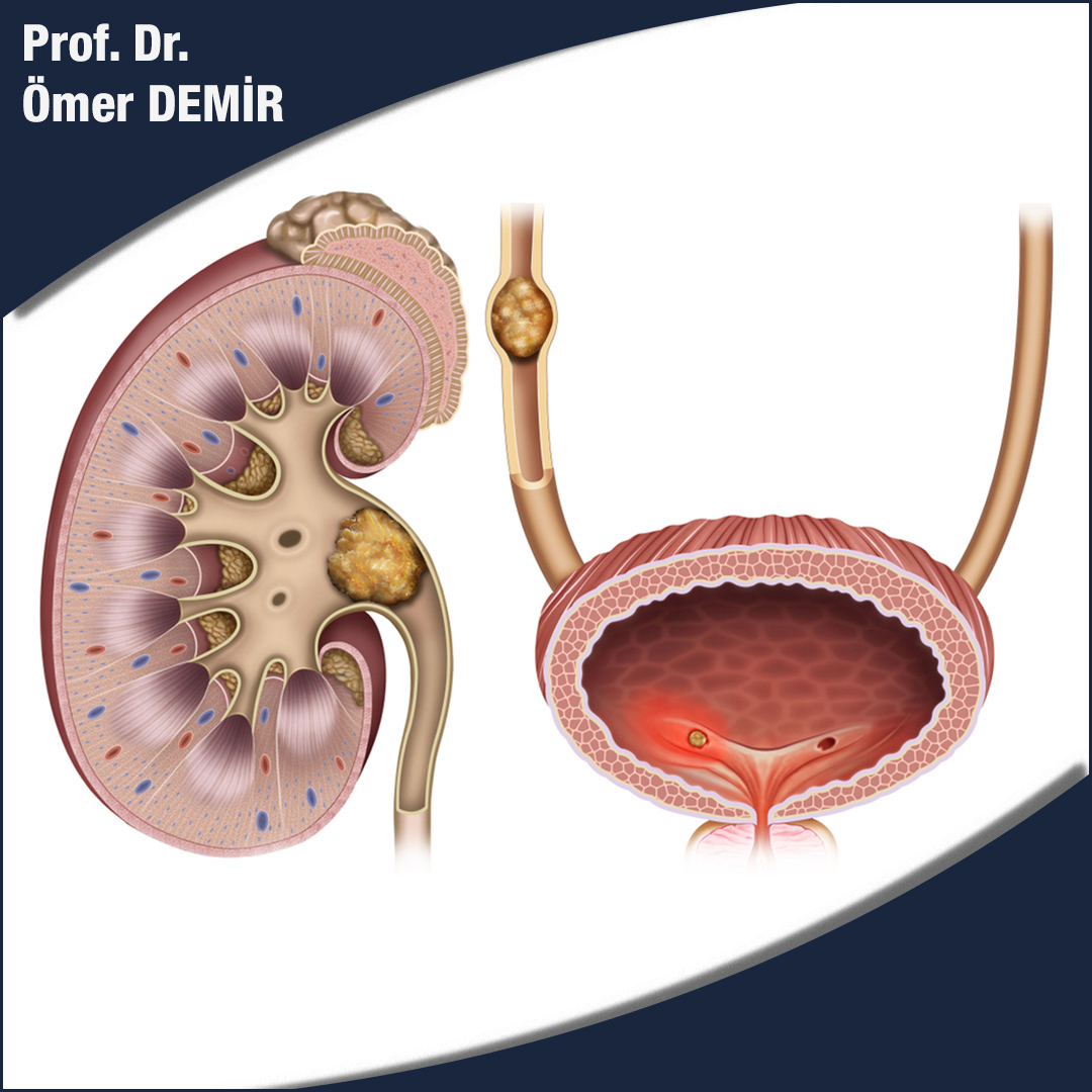
Kidney stones form when urine is supersaturated with salts and minerals such as calcium oxalate, struvite (ammonium magnesium phosphate), uric acid, and cystine. 60-80% of the stones contain calcium.
They can vary significantly in size, from small ‘gravel-like’ stones to large deer-horned stones. The stones may remain in the position they were formed or may travel in the urinary tract and cause complaints.
Bladder Stones
Bladder stones make up about 5% of urinary tract stones and are usually caused by foreign bodies, obstruction or infection. The most common cause of bladder stones is urine residue due to inability to empty the bladder completely while urinating, and most cases occur in men with bladder outlet obstruction.
Patients with permanent catheters are also at high risk of developing bladder stones, and there appears to be a significant relationship between bladder stones and the formation of malignant bladder tumors in these patients.
Frequency
Kidney stones are common, although most of them do not cause any complaints, they occur in one in ten people at some time in their life. It is 3 times more common in men than in women. Although urinary system stone disease can be seen at any age, it is most common between the ages of 30 and 50.
Risk factors
Various risk factors are known that increase the stone development potential of a susceptible individual.
These are:
1. Anatomical abnormalities of the kidneys and / or urinary tract – eg horseshoe kidney, ureteral stricture.
2. Family history of kidney stones.
3.Hypertension and diabetes
4. Gut.
5. Hyperparathyroidism.
6. Obesity and a sedentary lifestyle.
7. Lack of fluid intake.
8. Some metabolic disorders: chronic metabolic acidosis, hypercalciuria, hyperuricosuria.
9. Citrate deficiency in urine.
10. Some congenital metabolic diseases called cystinuria.
11. Medications (eg diuretics such as triamterene and calcium / vitamin D supplements).
12. It is more common in hot climates.
Complaints That May Occur
Most of the stones are asymptomatic and are noticed during examinations for other conditions. The classic feature of pain called renal colic is sudden severe pain. It is usually caused by stones in the kidney, kidney collecting structures or ureter and is caused by enlargement and stretching of the ureter. The pain begins in the lower back and flank (but sometimes lower) and may spread to the groin. The pain is usually accompanied by bleeding in the urine (hematuria). A moving stone is usually more painful than a stationary stone. Pain due to the stone at the lower end of the ureter usually spreads to the anterior part of the thigh, to the testicle and scrotum in men, and to the vagina in women. Symptoms such as nausea and vomiting, fever, burning in the urine, bleeding in the urine, and inability to urinate may accompany the pain.
Diagnostic Tests
Urine test; to look for infection and / or the presence of blood cells in the urine.
Blood test; to detect the presence of infection and kidney function.
Low-radiation drug-free computed tomography
Ultrasonography; In cases where computed tomography cannot be performed, especially to see kidney stones and enlargement in the kidney.
In patients with recurrent stone disease, special urine analysis can be performed by collecting 24-hour urine after the stone is cleaned.
Treatment
Approximately 1 out of 5 stones will not fall by itself and will require some kind of intervention. Especially stones that do not cause severe pain and obstruction can be expected to decrease with the use of abundant fluid intake, painkillers and drugs that dilate the urinary tract. The waiting period should not be longer than 6 weeks.
Procedures for removing stones include:
Extracorporeal shock wave lithotripsy (ESWL): ultrasonic shock waves are directed onto the stone to break up the stone. The stone particles are then expected to fall spontaneously with abundant water consumption.
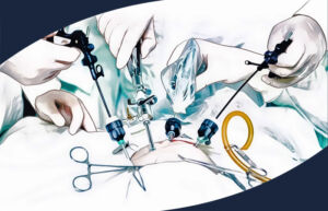
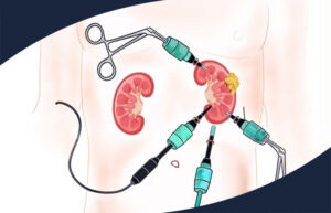
Percutaneous nephrolithotomy (PCNL): Used for large stones (> 2 cm), deer antler stones and also cystine stones. During the procedure, the kidney collecting system is entered through an approximately 1 centimetre incision from the flank using a device called a nephroscope. Stones are broken into pieces and removed using special stone crushing devices.
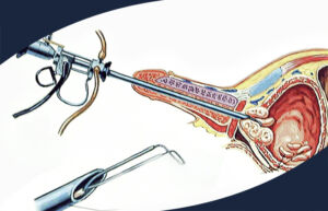
Ureteroscopy: It is a surgical technique applied for the treatment of stones in the ureter. Stones are broken and cleaned using a laser.
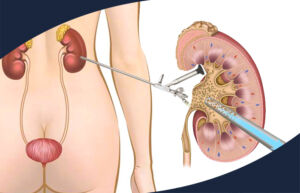
Retrograde Intrarenal Surgery (RIRS): Used for the treatment of stones smaller than 2 cm in the upper part of the ureter or kidney. In this surgical technique, laser is used to break up stones.
Open surgery: rarely required and usually applicable for complex cases or those who have failed all of the above.
Prevention of Stone Formation
Recurrence of urinary tract stones is common and therefore a variety of lifestyle measures should be recommended to help prevent or delay recurrence, especially after treatment of existing stone:
Increase fluid intake to keep urine output 2-3 liters per day.
Reduce your salt intake.
If you are obese, lose weight.
Reduce your consumption of meat and animal protein.
Reduce your intake of oxalate (oxalate-rich foods chocolate, coffee, nuts).
Keep calcium intake at normal levels (decreasing intake increases calcium oxalate excretion).

