PROSTATE CANCER
Prostate cancer is the most common type of cancer in men. It ranks second in cancer-related deaths.
Prostate cancer occurs when some cells that make up the prostate tissue show an abnormal course and form a tumor.
The tumor sometimes occurs in one part of the prostate, sometimes in more than one place. It can progress without causing any symptoms, and if it is not treated, it can grow and progress over time and cause urination disorders, bleeding in the urine or kidney failure.
Today, most of the patients diagnosed are diagnosed in the early period and long-term survival is achieved with effective treatments.
WHEN SHOULD WE SUSPECT PROSTATE CANCER?
The most important findings that make us suspect prostate cancer are that we persistently see results above normal values as a result of the PSA test and / or obtain suspicious findings in digital rectal examination.

Rectal Examination
Rectal examination, known as an anal finger prostate examination, is the palpation of the outer part of the prostate by the doctor to detect some abnormalities such as stiffness and irregularity in the prostate.
PSA (Prostate Specific Antigen) Test
Prostate Specific Antigen (PSA) is an enzyme produced in the prostate that regulates the density of the semen. A very small amount of PSA produced circulates in the blood. PSA passes into the blood in varying amounts in diseases such as benign enlargement of the prostate, prostate infections and prostate cancer. The PSA level in the blood and the change in this level can give an idea about the nature of prostate diseases. The frequency of PSA secretion is very important in diagnosing cancer. When you look at a normal prostate, it is seen that the PSA level in the blood is low. However, the low PSA value in the blood does not mean that the patient does not have prostate cancer. PSA value may be low in some local prostate cancers. The tumor can grow while at these levels. Determining the PSA value in the blood is very important for early diagnosis of prostate cancer and treatment as soon as possible.
DIAGNOSIS IN PROSTATE CANCER
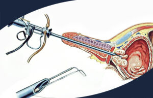
In cases where prostate cancer is suspected, the standard method used for diagnosis is prostate biopsy. The parts taken as a result of prostate biopsy are examined by pathology specialists for the presence of cancer and the result is reached. Biopsy can be done under local / general anaesthesia. During the procedure, an ultrasound probe is inserted into the rectum from the anus region of the patient and prostate images are taken. While imaging the prostate with ultrasound, 10-18 pieces are taken from certain areas and suspicious areas, taking into account the prostate size and patient characteristics. The most common side effects after the procedure are bleeding from the anus and urinary tract infections.
Biopsy can be performed with standard methods, and there are various methods to increase diagnostic efficiency. With the MR-fusion biopsy method performed under the guidance of MR images, the previously taken MR images are combined with the ultrasound device and special software, allowing biopsy from the suspicious areas seen here. In this way, a certain increase in the diagnostic efficiency of the biopsy procedure can be achieved.
TREATMENT IN PROSTATE CANCER
Treatment options in prostate cancer are determined by the stage of the disease at the time of diagnosis. Prostate cancer surgery (radical prostatectomy) is primarily recommended for patients with early diagnosis of prostate cancer in terms of cancer control and patient survival. Other treatment options that can be recommended considering the patient-related factors are various follow-up protocols and radiotherapy. Patients with advanced stage (metastasised prostate cancer) are followed up with sequential hormonal treatments and various chemotherapy protocols, and cancer patients can survive relatively longer when compared to other types of cancer.
Prostate cancer surgery (Radical prostatectomy)
In this surgery, seminal vesicles together with the prostate are completely separated from the urinary bladder (bladder) and urinary channel (urethra) and taken out of the body and sent for pathological examination. When necessary, the lymph nodes in the close area are surgically removed and pathological examination is performed. The remaining bladder and urethra are sutured close to each other through a catheter and the operation is completed. The main goal with this surgery is to provide cancer control and enable patients to live a longer life. However, after prostate cancer surgery, some changes may occur in patients’ lives and they may feel anxious. Most of the patients worry about situations such as urinary incontinence / incontinence in the postoperative period, but most of them (more than 95%) fulfill the urine retention function in the 3rd month of the operation. In other words, more than 95% of the patients do not have urinary incontinence problems. Yet another concern is erection problem. However, studies and experience have shown that in the preoperative period, young patients with completely normal erectile function and no additional risk factors can have an erection rate of 60-70% within 1 year after surgery.
Prostate cancer surgery can be performed with 3 different techniques today:
Open surgery (a single 10-12 cm incision under the navel)
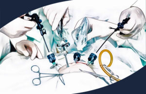
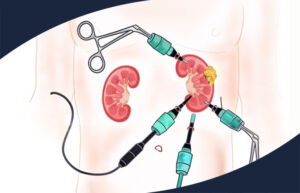
Laparoscopic surgery (5-6 incisions of 1-2 cm in the sub-umbilical region)
Robotic surgery (5-6 incisions of 1-2 cm in the sub-belly area)
Although these 3 techniques have their own advantages and disadvantages, each technique has no superiority to each other in terms of cancer control, urinary retention capability and recovery of erectile function.

